
My mentors at USC were Dr. Judy Pa and Vahan Aslanyan. Dr. Pa's lab is looking at early structural/functional changes and brain activity in older adults at risk for Alzheimer's disease. Their primary goal is to identify the earliest signs of cognitive decline and they hope to do that by combining cognitive, behavioral, and neuroimaging techniques. Since they are still in the early stages of the project, I assisted the team by looking at the Long beach Longitudinal Aging Study which investigated gender and APOE status differences in the relationship between brain health and risk measures. The team was still in the early stages of this project so there were no preliminary findings. The work I did during the summer was assisting the team with various assignments such as data manipulation, literature reviews, learning how to smooth out structural T1 magnetic resonance imaging (MRI) scans using VBM. Once the datasets are collected and imaging/sociodemographic data are combined, the team will then decide which area(s) of interest they plan to run statistical analyses on depending on the abundance of data they have. Right now, the lab is currently focused on exploring gender differences, so they expect to check for the significance of gender interactions with their variable of interest. Covariates such as age, APOE4, and education are included.
The USC Summer Research Experience taught me how to use various programs to work on data manipulation and examine MRI scans. I believe that this skill set I learned will be very useful in the future because Public Health gathers data from many studies and that data needs to be organized and examined by the scientific community and the public. It took some time to learn how each program worked and eventually I became familiar with them and create codes that assist me in data manipulation/harmonization. The experience also showed me that I can continue to improve my communication skills (such as presenting) because there is a lot of information/work that I want to convey to my audience, so I need to find a way to deliver it smoothly. I also learned how to prepare a presentation in a way that does not have too many slides and shows too much. Dr. Pa explained to me that depending on the time I have to present, that should be the number of slides I have as well. The advice/comments she gave me were very helpful and definitely something I will keep in mind for future presentations.
My mentor’s were Dr. Tyler Ard, Assistant Professor Of Research Neurology. Using data from the journal which study aims to present high resolution black blood MRI (2-D) for the visualization and characterizations of Lenticulostriate Arteries, it was my job to come up with the best way to visualize and represent these arteries in 3-D. The main objective was to see the structure of the Lenticulostriate Arteries and to get a better perspective on where they are located in the brain. Using a gaming developer software called Unity, I was tasked to create an interactive webpage of a human brain that highlighted these arteries. Preliminary findings included: In the United States, about 77.9 million (1 out of every 3) adults have high blood pressure and within that group about 8 out of 10 people will have at least one stroke. Cause of strokes are due to lack of oxygen to the brain. This lack of oxygen is caused by little to no blood flow to certain areas of the brain. No blood flow is due to blood clots which clogs already narrowed arteries. This leads to our Lenticulostriate arteries which are small and very susceptible to these blood clots. The use of Unity can help people understand the structure of Lenticulostriate Arteries and could possibly reduce the number of strokes.
I have experienced coding in C++ and working with GUI apps, but have never used the program called Unity. Unity is a great blend of C++ coding and working with 3-D models. I felt a bit intimidated, but I was looking forward to the challenge. I was able to experiment with my code and receive instant feedback. The project with Unity has helped me gain more confidence in my coding and understanding that this is all part of the learning experience. From this experience, I take with me the idea that even if I'm not sure of what I am doing, I know I have the tools to get me there.
My mentor at USC was Dr. Hosung Kim, Assistant Professor of Neurology. His main areaf o research is neuroimaging with deep learning. The main research objective was to see Parkinson's data set on current brain age prediction algorithm to predict brain age of Parkinson's patients. We hypothesized that the brain age would be accelerated for patients of Parkinson's disease. Our preliminary findings show showed that brain age was not accelerated in the patients of Parkinson's disease dataset. Some implications are that parameter tuning may help to prevent potential over-fitting of the dataBrain age prediction algorithm can be run on various subsections of the brain to observe if parts of the brain in Parkinson's patients is accelerated more than other parts. Parts of the brain most affected by Parkinson’s (substantia nigra, motor cortex) may show anomalies in brain aging.
Working in the lab was fun. I want to understand which models are appropriate to use with different kinds of data. Communication is important in the lab. If you're having trouble finding something, you should speak up instead of wasting time. I used Pintask to keep track of objectives and to document steps in processes. It was a good tool to use to help stay on track with time. (Good when you're working by yourself). I learned that my skills are needed and that I know more than I was giving myself credit for.
My mentor was Dr. Tyler Ard, Assistant Professor of Research, and his main area of research: Visualizing neuroimaging data. Our main research objective was to develop a program capable of removing noise from images taken from two photon microscopy so that a stack of images can be modeled in three dimensions. Preliminary findings: Noise data in images can be removed using methods such as windowing, tuning threshold parameters, and blurring.
Next steps/conclusions: Removing noise from images was successful, however the same processing stack will need to be tested on other stacks of images including those taken from methods that do not include two photon microscopy. Prior to starting my summer at USC, I had no prior experience with image processing. Within a nine week period I was able to learn about an array of methods to process images including methods that I created entirely on my own. I effectively learned how to process images having had no prior knowledge in a short time period.
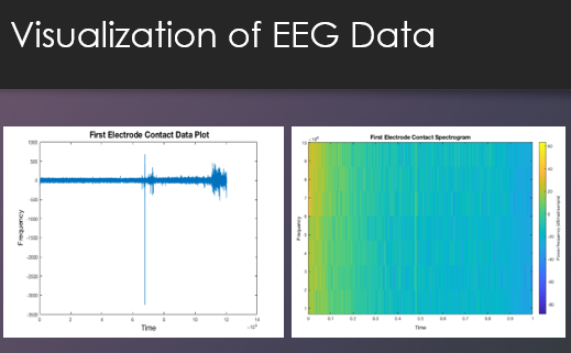
My mentor was Dr. Jann who is currently an Assistant Professor at the USC Stevens Institute for Neuroimaging and Informatics. Dr. Jann has a strong background in multimodal imaging, combining electrophysiological, functional and structural MRI methods. His research focuses on developing novel analysis approaches to delineate the brain’s functional organization in health and disease.
The title of my project was: The Relationship Between Cognitive Function and Complexity of functional magnetic resonance imaging (fMRI). My research focused on cognitive performance by analyzing the multi-scale entropy of nodes within a network related to working memory and executive function. Future research may lead to predicting cognitive decline by focusing on the complexity of each node. It may be possible to help us determine when a network will fail based on the performance of the nodes that form part of that network. I had a great experience at USC! I learned so much about the complexity of the brain and how much we still need to learn about it. I learned how to code using Matlab. Working with Dr. Jann helped me to experience how the research process works.
At USC, Melissa investigated the effect of Alzheimer's and dementia on brain complexity by analyzing MRI brain scans. During this project, she had the opportunity to gain experience programming in MATLAB and using data visualization tools to better understand brain maps.
Nayelie had the opportunity to spend her summer at USC to work in the lab of Dr.Braskie, a neuroscientist. The efforts of Nayelie were to assist Dr.Braskie’s on her research on Alzheimer’s amongst the Mexican- American community. She had the opportunity to learn to code on shell with bash which allows for stripping of the skulls from MRI images; the brain data is going to be used for analysis. Her experience had an influence on her in regards to the future possibility of doing research on how chronic diseases affect brain health.
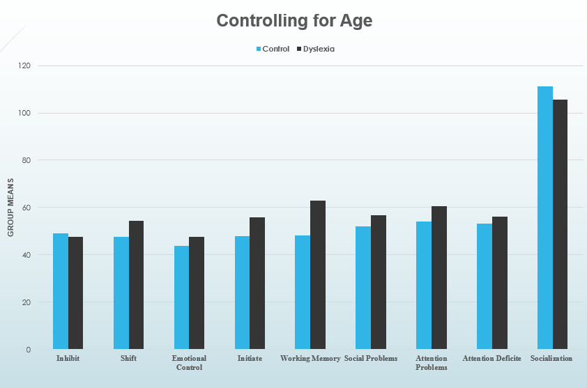
My research at the Mark and Mary Steven’s Neuroimaging and Informatics Institute involved a sample of children with dyslexia. These children have been scanned using MRI, to further understand these issues at a neurobiological level. My project looks at the differences in behaviors between dyslexic children and controls, specifically in social skills and attention. Additionally, I analyzed brain structures thought to participate the functioning of these variables; However, differences were only noticed with variables related to attention, such as working memory, shifting, and initiation, while no significant differences were found in social skills. Therefore, neuronal analysis on areas related to socializing were not pursued. Neuronal analysis involved comparing the surface area, gray matter volume and average cortical thickness between the two group. Intra-cranial volume was controlled for in the brain analysis. The lateral prefrontal cortex, dorsal anterior cingulate cortex, and posterior parietal lobe, areas thought to participate in attention, were the focus of the neuronal analysis. Results the brain analysis did not reveal any significant differences between dyslexic children and controls, likely meaning that structure size is not related to the problems that manifest in dyslexia. The purpose of this research was to better understand the nature of the issues that manifest in dyslexia.![]() Download: Dyslexia Presentation
Download: Dyslexia Presentation
Mentored with Dr. Dominique Duncan in an international and multidisciplinary study named the Epilepsy Bioinformatics Study for Antiepileptogenic Therapy (EpiBioS4Rx). The study is aiming to create cost effective and full-scale clinical trials for the prevention of epilepsy. Additionally, the study is working to identify biomarkers for epileptogenesis using the big data generated across the study's centers.![]() Download: Epilepsy Bioinformatics Presentation
Download: Epilepsy Bioinformatics Presentation
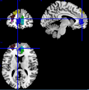
Functional magnetic resonance image (fMRI) data was used from the Human Connectome Project 900 Subject Data set. The goal of my project was to identify brain regions that may be connected to each other. An atlas was used to split up the gray matter of the brain into 200 different nodes. With the use of Matlab and Unix commands, I created a matrix and performed clustering on the 200 brain nodes. As a result, this provided 20 different clusters. Each cluster included brain nodes that were identified as having a statistically similar functional brain connectivity pattern. The clusters were then transferred onto a software, MRIcron, where it showed what brain nodes were connected to other brain nodes. I was able to successfully identify highly consistent brain networks such as the default mode network and sensory network. ![]() Download: Brain Connectivity Analysis Presentation
Download: Brain Connectivity Analysis Presentation
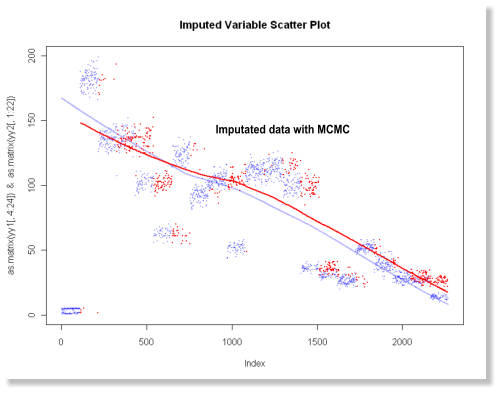
The current study on neurodevelopment is conducted as part of Dr. Kristi Clark’s lab at USC, with mentorship from Dr. Farshid Sepehrband. Understanding the nuances of the brain in its development has far-reaching implications for research and clinical practice. The field of neurodevelopment has been examined using methods such as MRI and fMRI scans, and research in the field can allow us further understanding of the growth of the brain and central nervous system. Advances in computer science and data storage methods allow researchers to collect, store, and ultimately analyze the large neuroimaging datasets. The Philadelphia Neurodevelopment Cohort (PNC) study is one such example of the efforts made in examining neurodevelopment, with over 1000 participants imaged. See more at: Data Imputation Techniques